Human cornea harbors tissue-resident memory T cells shaped by systemic immune activation, age and biological sex

Human cornea harbors tissue-resident memory T cells shaped by systemic immune activation, age and biological sex
Wu, M.; Monard, S. C.; Yakou, M. H.; Senthil, K.; Devi, S.; Alexandre, Y. O.; Wu, C. Y.; Burn, T. N.; Groom, J. R.; Nguyen, B. N.; Mackay, L. K.; Chinnery, H. R.; Mueller, S. N.; Downie, L. E.
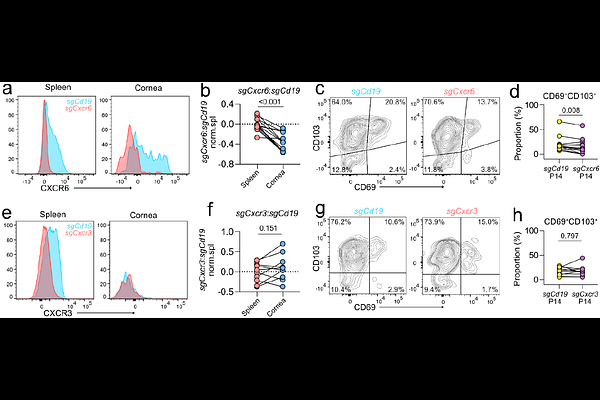
AbstractThe cornea is classically regarded as an immune-privileged tissue. However, recent studies have identified T cells in the healthy human cornea that are absent in specific pathogen-free (SPF) mice. The identity of these T cells, the systemic cues that drive their establishment, and the molecular mechanisms governing their corneal homing remain unknown. Here, we combine multimodal human data with tractable murine models to characterize the cellular basis of corneal immune surveillance. Immunofluorescence staining and confocal imaging of clinically non-inflamed human donor tissues revealed CD3 T cells within the corneal epithelium. Flow cytometry revealed that these cells were predominantly CD8, with a tissue-resident memory T (TRM) cell phenotype. In vivo confocal microscopy demonstrated that corneal T cell abundance increased with age, particularly in males, suggesting that accumulation is shaped by cumulative systemic immune experience. Whereas young SPF mice were devoid of corneal T cells, infection with pathogens that do not typically target the cornea induced long-lived TRM-like CD8 T cells in the cornea. We identified CXCR6 as required for efficient T cell recruitment to the cornea of viral-infected mice. Together, our human and murine data support a model in which systemic immune history, age and biological sex influence local immune surveillance at the human ocular surface.