Proliferation of Early and Mature Osteoblasts Is Required for Bone Fracture Healing in Mice
Proliferation of Early and Mature Osteoblasts Is Required for Bone Fracture Healing in Mice
Gould, N. R.; Coello, A. F.; McKenzie, J. A.; Li, T.; Hixon, K. R.; Chen, L.; Barwick, K.; Lee, T.; Obaji, M.; Zhang, B.; Ornitz, D. M.; Silva, M. J.
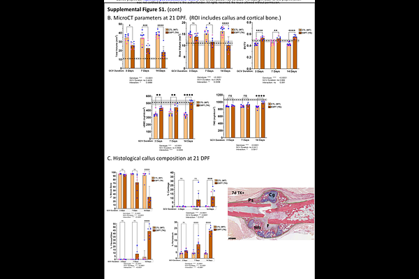
AbstractFractures heal by rapid formation of mineralized callus. Essential to this process is proliferation of periosteal cells to supply bone-forming osteoblasts. To better understand the role of cell proliferation in fracture healing, we asked: How is callus composition altered when osteolineage cells proliferation is blocked? Do mature osteoblasts proliferate to contribute to callus formation? First, mice expressing herpes simplex virus-thymidine kinase (HSV-TK) driven by the 3.6Col1a1 promoter were treated with ganciclovir (GCV) to ablate proliferating osteolineage cells for 5 or 10 days. Analysis of callus cells using single-cell RNA-seq revealed that GCV-treated Col1-TK mice had fewer osteoblasts and chondrocytes than control mice, with more myofibroblasts and immune cells, consistent with fibrous nonunion. In controls, 15-30% of callus cells that expressed the early osteoblast markers osterix (OSX) and the late marker osteocalcin (OCN) were in the cell cycle. Next, we targeted proliferating osteoblasts at different stages of differentiation by crossing Osx-CreERT2, Ocn-Cre and Dmp1-CreERT2 mice with novel ROSA-TK mice. Following fracture, each Cre;ROSA-TK mouse line exhibited poorer radiographic healing, decreased callus bone volume and a shift from callus bone to fibrous tissue. We conclude that osteoblasts, often considered post-mitotic, proliferate after fracture to contribute to formation of mineralized callus essential to healing.