Intermingled representation of oral cavity in mouse trigeminal ganglion

Intermingled representation of oral cavity in mouse trigeminal ganglion
Matunis, A.; Iwamoto, R.; Stacy, E.; Abe, K.; Tamura, S.; Kambe, Y.; Itokazu, T.; Hikida, T.; Sato, T.; Sato, T.
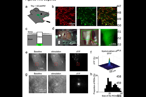
AbstractSomatotopy serves as a fundamental principle underlying sensory information processing, traditionally emphasized in the study of the cerebral cortex. However, little effort has been directed towards unraveling the spatial organization characterizing the earlier stages of somatosensory pathways. In this study, we developed a novel methodology to visualize individual neurons within the trigeminal ganglion, a crucial cluster of cell bodies of sensory neurons innervating the face. Our investigations revealed a reliable sensory response to stimulation of the lower incisor or lip within this ganglion. The responsive neurons were confined to a specific portion of the trigeminal ganglion, consistent with innervation of the lower oral cavity by the mandibular nerve (V3). Contrary to our expectations, we did not observe a discernible map differentiating the lower tooth and the lip. Instead, the spatial representation of the tooth and lip within this portion of trigeminal ganglion exhibited intermingling with no clear border between tooth-responding and lip-responding neurons. These findings contrast with earlier studies that identified tooth and lip responding regions in the somatosensory cortex. Our study sheds light on the complex spatial organization of sensory processing in the trigeminal system, highlighting the need for further research to elucidate the underlying mechanisms and implications for sensory perception and clinical interventions.