Multimodal mechano-SICM and FRET with stretch for probing cardiomyocyte function.
Multimodal mechano-SICM and FRET with stretch for probing cardiomyocyte function.
Reilly-O'Donnell, B.; Shevchuk, A.; Gorelik, J.
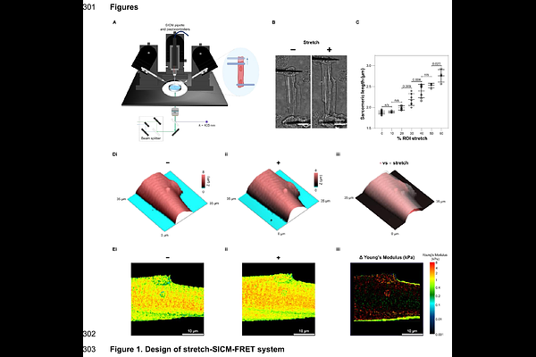
AbstractCardiac function is dependent upon the ability of cardiomyocytes to adapt their contractions to meet the demands of the body. Increased preload lengthens the sarcomere, altering the efficiency of contraction. The surface topography of cardiomyocytes is distinct from other cell types. T-tubules are key membrane structures which protrude into the cell body, aligned with the edges of the sarcomere. These are key signalling domains which ensure efficient and adaptable excitation-contraction coupling. It has been shown that T-tubules are dynamic structures which deform during the contraction cycle however, how the T-tubule structure adapts to increased preload has not been realised. Here we demonstrate a methodology for the measurement of the surface topography and sub-cellular signalling of isolated adult cardiomyocytes under diastolic stretch. We track individual T-tubule openings, showing that increased load causes them to shift, increase diameter and become stiffer. Future applications of this system include experimental modelling of preload-reducing therapies, for the treatment of acute and chronic heart failure.