Exploring non-invasive sexing of early chick embryos in intact eggs using Laser Speckle Contrast Imaging (LSCI) and Deep Neural Network (DNN)
Exploring non-invasive sexing of early chick embryos in intact eggs using Laser Speckle Contrast Imaging (LSCI) and Deep Neural Network (DNN)
Mahler, S.; Arora, A.; Readhead, C.; Yin, S.; Hari, S. N.; Wang, E.; Moxley, C. I.; Adeboye, A. A.; Dong, Z.; Zhou, H.; Chen, X.; Bronner, M.; Yang, C.
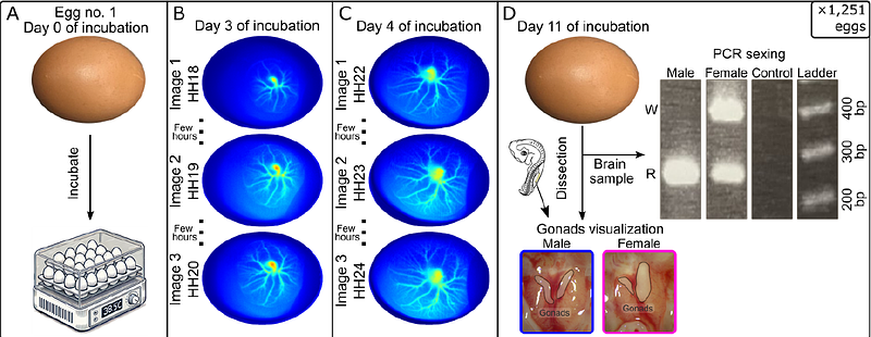
AbstractThe ability to image blood flow in early-stage avian embryos has significant applications in developmental biology, drug and vaccine testing, as well as determining sex differentiation. In this project, we used our recently developed laser speckle contrast imaging (LSCI) system to non-invasively image extraembryonic blood vessels and used these images to attempt early sex identification of chick embryos. Specifically, we captured images of blood vessels from 1,251 living chicken embryos between day three and day four of incubation. We then applied deep neural network (DNN) models to evaluate whether it is possible to differentiate sex based on vascular patterns. Using ResNetBiT and YOLOv5 models, our results indicate that sex differentiation from extraembryonic blood vessel images was not achievable with sufficiently high accuracy or statistical significance for practical use. Specifically, ResNetBiT had a five-fold cross-validated average accuracy of 59%{+/-}5% (fold-wise p-value, p [≤] 0.3) at day 3 and 61%{+/-}3% (fold-wise, p [≤] 0.04) at day 4. YOLOv5 had a five-fold cross-validated average accuracy of 55%{+/-}3% (fold-wise, p [≤] 0.3) at day 3 and 53%{+/-}3% (fold-wise, p [≤] 0.5) at day 4. Our findings suggest that using vascular pattern imaging alone is inconclusive for reliable early sex identification in chicken embryos.