sPRR signaling in macrophages via the AT1R/Yap/Taz axis to induce renal fibrosis
sPRR signaling in macrophages via the AT1R/Yap/Taz axis to induce renal fibrosis
Feng, Y.; Zheng, H.; Xie, S.; Wang, F.; Luo, R.; Yang, T.
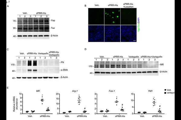
AbstractBackground: Within the kidney, (pro) renin receptor (PRR) is abundantly expressed in the collecting duct (CD) and modulate physiological and pathophysiological processes. We have recently reported that activation of CD PRR mediates obstructive renal fibrosis in a mouse model of unilateral ureteral obstruction (UUO). The current study addresses the underlying mechanisms by examining the profibrotic pathway mediated by soluble PRR (sPRR)-dependent alternative macrophage activation. Methods: We performed UUO or sham surgery on mice with CD-specific deletion of PRR (CD PRR KO) and floxed controls. After 7 days, we assessed fibrosis-related parameters, inflammatory cytokines, M1/M2 macrophage markers, other gene expression markers of kidney injury, and the concentration of plasma sPRR. We also administered vehicle or site-1 protease (S1P) inhibitor PF-429242 (PF) to C57BL/6 mice with UUO to determine the role of sPRR. Experiments were performed in vitro to examine the mechanism of sPRR-His-mediated macrophage M2 polarization and activation of potential target genes in bone-marrow-derived macrophages (BMDMs). Results: Compared with the floxed control, CD PRR KO decreased macrophage accumulation, M2 polarization, and Yap/Taz expression while improving renal fibrosis and suppressing plasma sPRR levels following UUO. In BMDMs, sPRR-His treatment promoted macrophage M2 polarization, fibrosis, and Yap/Taz expression, which was dependent on angiotensin type 1 receptor (AT1R). Conclusion: CD PRR-derived sPRR acts via ATR to promote macrophage M2 polarization and stimulates the AT1R/Yap/Taz axis, which leads to renal fibrosis during UUO.