Plaque-associated Microglial Polarization in Visual Brain Regions of the 5xFAD Mouse Model
Plaque-associated Microglial Polarization in Visual Brain Regions of the 5xFAD Mouse Model
McCool, S.; Smith, J. C.; Van Hook, M.
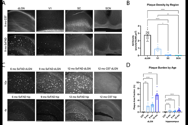
AbstractAlzheimer's disease (AD), a neurodegenerative disorder associated with amyloid beta (A{beta}) plaque deposition, leads to cognitive decline in affected individuals. Vision changes are some of the first reported symptoms in AD with studies showing both decline in functions performed by the visual system as well as associations between vision loss and cognitive impairment in AD patients. Due to the increasing number of individuals diagnosed with AD and its early impact on vision, we sought to provide an in-depth analysis using immunohistochemistry and 2-photon imaging techniques in the 5xFAD mouse model of amyloidosis to examine specifically how A{beta}, a primary pathology typically preceding many other AD-associated pathologies, affects visual regions of the brain and how microglia, key immune regulators of the brain's environment, respond to this AD-like pathology. We found that in the pathway for image-forming vision, including the dorsolateral geniculate nucleus (dLGN) and the primary visual cortex (V1), there was significant A{beta} pathology and shifts in microglial morphology to an amoeboid state and increased phagocytic activity. However, in non-image-forming visual brain regions such as the superior colliculus (SC) and suprachiasmatic nucleus (SCN), there was minimal A{beta} pathology, ramified microglial morphology, and minimal phagocytic activity. Overall, visual brain regions associated with A{beta} plaque deposition experience significant microglial polarization when examining both morphology and function.