Comparing Linear and Nonlinear Finite Element Models of Vertebral Strength Across the Thoracolumbar Spine: A Benchmark from Density-Calibrated Computed Tomography
Comparing Linear and Nonlinear Finite Element Models of Vertebral Strength Across the Thoracolumbar Spine: A Benchmark from Density-Calibrated Computed Tomography
Walle, M.; Matheson, B. E.; Boyd, S. K.
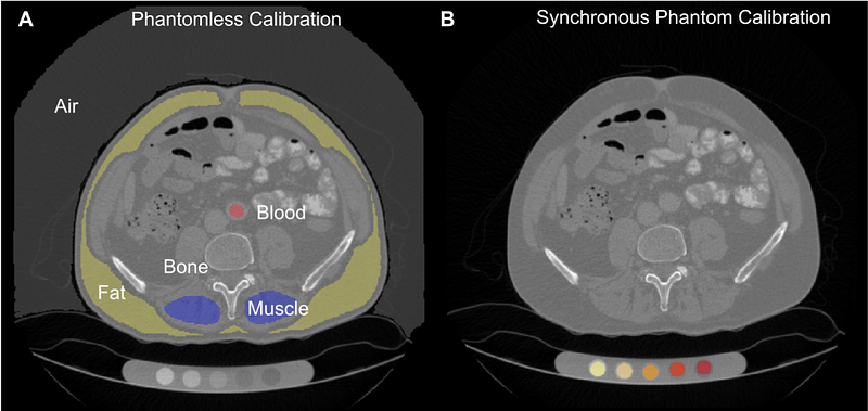
AbstractOpportunistic assessment of vertebral strength from clinical computed tomography (CT) scans holds substantial promise for fracture risk stratification, yet variability in calibration methods and finite element (FE) modeling approaches has led to limited comparability across studies. In this work, we provide a publicly available benchmark dataset that supports standardized biomechanical analysis of the thoracic and lumbar spine using density-calibrated CT data. We extended the VerSe 2019 dataset to include phantomless quantitative CT calibration, automated vertebral substructure segmentation, and vertebral strength estimates derived from both linear and nonlinear FE models. The cohort comprises 141 patients scanned across five CT systems, including contrast-enhanced protocols. Phantomless calibration was performed using automatically segmented tissue references and validated against synchronous calibration phantoms in 17 scans. To evaluate model performance, we implemented a nonlinear elastoplastic FE model and compared it to two linear estimates. A displacement-calibrated linear model (0.2% axial strain) demonstrated excellent agreement with nonlinear failure loads (R = 0.96; mean difference = -0.07 kN), while a stiffness-based approach showed similarly strong correlation (R = 0.92). We evaluated vertebral strength at all thoracic and lumbar levels, enabling level-wise normalization and comparison. Strength ratios revealed consistent anatomical trends and identified T12 and T9 as reliable alternatives to L1 for opportunistic screening and model standardization. All calibrated scans, segmentations, software, and modeling outputs are publicly released, providing a benchmark resource for validation and development of FE models, radiomics tools, and other quantitative imaging applications in musculoskeletal research.