Fetal sex shapes placental inflammatory responses to extracellular mitochondrial DNA

Fetal sex shapes placental inflammatory responses to extracellular mitochondrial DNA
da Silva, R. d. N. O.; Hula, N.; Escalera, D.; Lopez, L.; Kelly, G.; Gorham, I. K.; Rowe, M.; Ricci, C. A.; Gheorghe, C.; Phillips, N. R.; Goulopoulou, S.
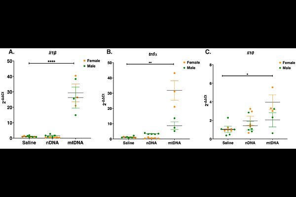
AbstractAberrant changes in circulating cell-free mitochondrial DNA (ccf-mtDNA) across gestation are associated with adverse pregnancy outcomes. Given the inflammatory properties of ccf-mtDNA via pattern recognition receptors such as Toll-like receptor 9 (TLR9), we hypothesized that extracellular mtDNA induces placental inflammation via TLR9 signaling and that this response differs by fetal sex. Pregnant Sprague-Dawley rats were treated intravenously with purified mtDNA (300 g/kg), nuclear DNA (nDNA), saline, and/or the TLR9 antagonist ODN2088 across five studies. Placental responses were evaluated 4 h (Studies 1-3) and 24 h (Study 4) post-treatment; pregnancy and neonatal outcomes were assessed at delivery (Study 5). Exposure to mtDNA, but not nDNA, increased placental il1{beta}, tnf, and il10 mRNA (p < 0.05), establishing response specificity. mtDNA-induced placental inflammation was fetal sex-dependent: mtDNA increased il6 and il1{beta} mRNA in male placentas (p [≤] 0.0004) but not female placentas, whereas ifn was selectively induced in female placentas (p = 0.0004). TLR9 and MyD88 abundance increased in female but not male placentas, and TLR9 antagonism modified selected inflammatory responses with sex-specific patterns. The 4 h inflammatory transcriptional signature resolved by 24 h, whereas mtDNA exposure was associated with a sex-specific shift in antioxidant enzyme expression persisting to 24 h. Despite no effects on gestational length or neonatal biometrics, mtDNA exposure was associated with a higher estimated stillbirth count per litter (IRR = 4.23, 95% CI [0.89, 20.1], p = 0.069). These findings establish extracellular mtDNA as an acute, sex-differentiated placental inflammatory stimulus with partial TLR9 dependence and a potential impact on fetal viability.