Lysosomal abundance in young and aged mouse hearts assessed by In Vivo Imaging Systems (IVIS) Lysotracker imaging and autophagy-related gene expression
Lysosomal abundance in young and aged mouse hearts assessed by In Vivo Imaging Systems (IVIS) Lysotracker imaging and autophagy-related gene expression
Albulushi, J.; Coghlan, H.; Moothanchery, M.; Dev, A.; Akerman, E.; Heenan, J.; Helassa, N.; Adegbite, O.; Sharma, P.; Patel, F.; Harrison, L.; Maguire, M. L.; Mirams, G. R.; Sweitach, P.; Poptani, H.; Burton, R. A. B.
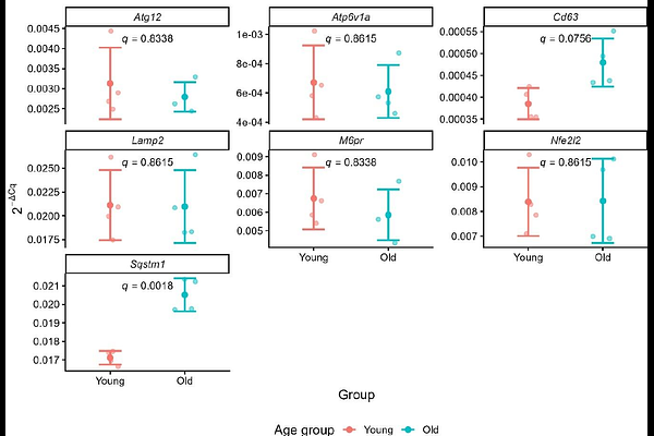
AbstractLysosomal function is essential for cardiac proteostasis and cellular health, yet its regulation during ageing remains poorly defined. We hypothesised that ageing alters both the abundance of acidic organelles and the machinery supporting their acidification. Using fluorescence-based In Vivo Imaging Systems (IVIS) with Lysotracker Red in young (2 to 4 months) and aged (18 months) mouse hearts, we quantified whole-heart acidic-vesicle signals and assessed expression of lysosomal and autophagy-related genes (Lamp2, Atp6v1a, Sqstm1, Cd63, Atg12, Nfe2l2, M6pr) by RT-qPCR. Whole-heart labelled Lysotracker fluorescence did not differ significantly between age groups, indicating preservation of the total acidic-vesicle pool. No changes in Atp6v1a and Lamp2 expression suggest acidification capacity and structural stability are maintained, whereas the minor, upregulation of Sqstm1 might indicate increased autophagic demand and altered vesicle trafficking, which warrants further investigation. No statistical significant changes in M6pr, Atg12, or Nfe2l2 were detected, suggesting transcriptional stability in enzyme trafficking, core autophagy, and oxidative stress pathways. Regionally, atria showed higher Lysotracker signal than ventricles, consistent with known enrichment of acidic vesicular stores in atrial physiology. These findings highlight the utility of IVIS imaging of Lysotracker-labelled hearts, providing rapid whole-organ assessment of acidic vesicle distribution, albeit with limited depth resolution. Complementary techniques such as RT-qPCR analysis is essential to interpret IVIS findings, enabling insight into underlying molecular changes in lysosomal and autophagy pathways during cardiac ageing.