Neuronal or vascular receptive fields? On the relation between BOLD-fMRI population receptive field (pRF) estimates and cortical vascularization.
Neuronal or vascular receptive fields? On the relation between BOLD-fMRI population receptive field (pRF) estimates and cortical vascularization.
Schellekens, W.; Nota, M.; Groen, I. I. A.; Piantoni, G.; Winawer, J.; Petridou, N.
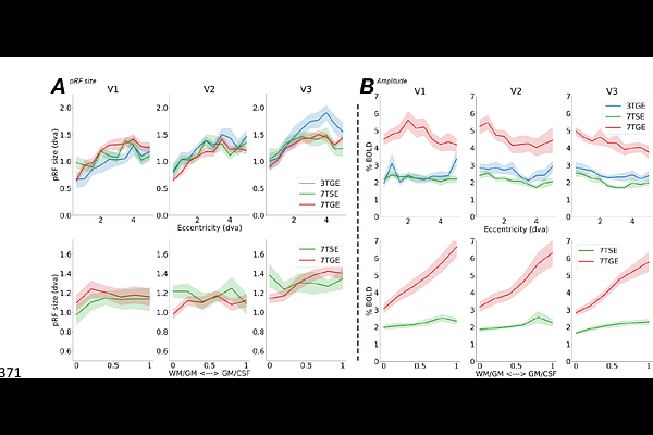
AbstractThis study investigates the contribution of different vascular compartments on population receptive field (pRF) size estimates within the early visual cortex (V1, V2, and V3) using BOLD-fMRI. We employed T2*-weighted gradient-echo (GE) and T2-weighted spin-echo (SE) sequences at 7 Tesla (7T) and a multi-band GE sequence at 3T to explore how different vessel sensitivities across these sequences influence pRF modeling. Our results confirm the expected pRF size increase across eccentricity and visual areas but found no significant differences in pRF size estimates across MR sequences, voxel sizes, or field strengths. BOLD signal amplitudes were influenced by MR sequence, with the largest signal changes observed for 7TGE, and amplitude increases were noted across cortical depth for GE sequences but not for SE sequences. Contrary to our hypotheses, pRF size estimates were not noticeably affected by the local vascularization, suggesting that pRFs primarily reflect neuronal activity rather than vascular compartment characteristics. Our study highlights the robust nature of pRF size estimates across various fMRI conditions and points toward the decoupling of pRF properties from vascular factors.