Fornix subdivisions and spatial learning: a diffusion MRI study
Fornix subdivisions and spatial learning: a diffusion MRI study
Hodgetts, C. J.; Postans, M.; Williams, A. N.; Graham, K. S.; Lawrence, A. J.
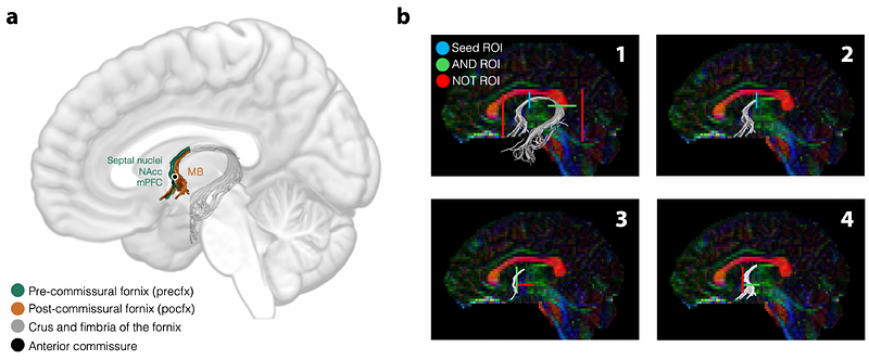
AbstractThe fornix is the major white matter tract linking the hippocampal formation with distal brain sites. Human and animal lesion studies show that the connections comprising the fornix are vital for specific attributes of episodic and spatial memory. The fornix, however, interconnects the hippocampal formation with an array of subcortical and cortical sites and it is not known which specific connections support spatial-mnemonic function. To address this, making use of a partly previously published dataset (Hodgetts et al., 2020), we applied a novel deterministic tractography protocol to diffusion-weighted magnetic resonance imaging (dMRI) data from a group of healthy young adult humans who separately completed a desktop-based virtual reality analogue of the Morris water maze task. The tractography protocol enabled the two main parts of the fornix, delineated previously in axonal tracing studies in rodents and primates, to be reconstructed in vivo, namely the pre-commissural fornix (connecting the hippocampus to the medial prefrontal cortex and the basal forebrain) and the post-commissural fornix (connecting the hippocampus to the medial diencephalon). We found that inter-individual differences in pre-commissural- but not, surprisingly, post-commissural- fornix microstructure (indexed by free water corrected fractional anisotropy, FA) were significantly correlated with individual differences in spatial learning, indexed by reduction in search error as individuals learned to navigate to a hidden target location from multiple starting points. This study provides novel evidence that flexible and/or precise spatial learning involves a hippocampal-basal forebrain/prefrontal network underpinned in part by the pre-commissural fornix.