Dynamics of ML-based Morphological Features Indicate a Shear Stress-Dependent Bifurcation of hiPSC-Derived Endothelial Cell States

Dynamics of ML-based Morphological Features Indicate a Shear Stress-Dependent Bifurcation of hiPSC-Derived Endothelial Cell States
Angelini, E.; Leveille, C. L.; Parent, S. E. P. E.; Zaunbrecher, R. J.; Barszczewski, T.; Dixon, J. C.; Mohammed, F. S.; Morris, B.; Yu, J.; Arakaki, J.; Dupar, R. J.; Edmonds, J. H.; Ehlers, E. A.; Gamlin, C. R.; Hedayati, M. J.; Hookway, C.; McCarley, J.; Mogre, S. S.; Phan, A.; Roberts, B.; Sanchez, E. E.; Thottam, J. P.; Wijesooriya, C. S.; Yao, J.; Kutys, M. L.; Nazockdast, E.; Wang, J.; Theriot, J. A.; Dalgin, G.; Rafelski, S. M.; Viana, M. P.
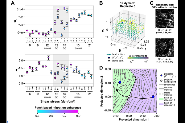
AbstractCell states are increasingly conceptualized as attractors of high-dimensional dynamical systems, yet quantitative approaches for integrating phenotypic information into this framework remain limited. Here, we take an image-based approach that combines unsupervised machine learning (ML) with timelapse imaging to extract and characterize the temporal dynamics of morphological features. Using a cell line with endogenously tagged VE-cadherin, we acquired brightfield and fluorescence timelapse images of human induced pluripotent stem cell-derived endothelial cell (hiPSC-EC) monolayers, which adopt distinct phenotypes at two different magnitudes of shear stress in terms of their morphology, behavior, and VE-cadherin organization. To quantify these phenotypic cell states without segmentation, we trained a diffusion autoencoder to predict VE-cadherin signal from brightfield images. We identified interpretable ML-based features representing cell orientation, elongation, and local density. Treating these variables as dimensions of a morphological state space, we estimated a data-driven vector field and found that the two observed phenotypic cell states correspond to stable fixed points of the inferred dynamical system. Mapping measured cell migration coherence onto this space further distinguished the states. Imaging cells across intermediate shear stresses revealed a regime of bistability in which both states coexist, indicating that the shear-stress-dependent transition between endothelial cell states occurs as a bifurcation of the inferred dynamical system. Finally we applied this method to study an N-terminal truncation of VE-cadherin, finding that mutated cells preserve alignment and coherent migration, but exhibit altered morphology and increased migration speed. This work demonstrates the applicability of a dynamical systems approach to quantitatively characterize morphological aspects of cell state from interpretable ML-based features.