Differential effects of fenofibrate and fenofibric acid on the regulation of liver endothelial permeability
Differential effects of fenofibrate and fenofibric acid on the regulation of liver endothelial permeability
Luty, M. T.; Borah, D.; Szafranska, K.; Giergiel, M.; Trzos, K.; McCourt, P.; Lekka, M.; Kotlinowski, J.; Zapotoczny, B.
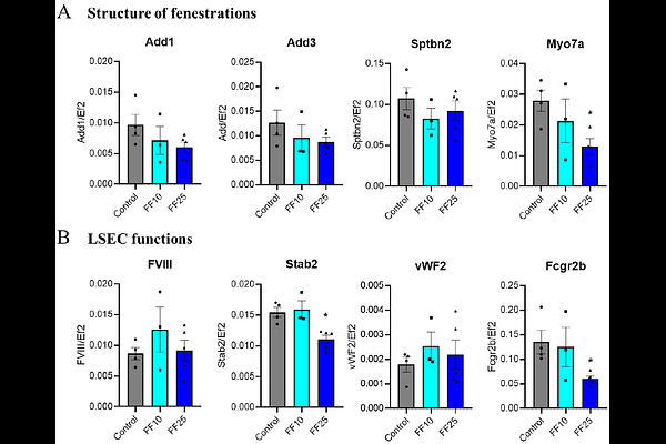
AbstractBackground and Aims: Fenofibrate is widely prescribed for hyperlipidaemia and has been associated with rare but severe cases of drug-induced liver injury (DILI), yet its effects on liver sinusoidal endothelial cells (LSECs) remain to be investigated. LSECs maintain a highly permeable specialized sinusoidal barrier characterized by transcellular pores (fenestrations), regulating the bidirectional transfer of circulating compounds to and from the hepatocytes. As drug-induced alterations in fenestration architecture could influence xenobiotic access to hepatocytes, these changes may modulate pathways associated with DILI. Understanding the effects of fenofibrate on LSEC ultrastructure may therefore provide insights into previously underexplored endothelial contributions to hepatic drug responses. Methods: Both fenofibrate and its active metabolite, fenofibric acid, were evaluated for their effects on LSEC ultrastructure, mechanical properties, and functional markers. Atomic force microscopy (AFM) and scanning electron microscopy (SEM) and were used to quantify fenestration architecture. AFM was additionally used to measure cellular mechanical properties, which were interpreted in the context of fluorescence-based quantification of cytoskeletal organization. Gene expression, viability, and cytotoxicity were assessed using PCR based and biochemical assays. Results: Fenofibrate reduced fenestration number and porosity at both tested concentration (10, and 25 uM). It also decreased the apparent Youngs modulus of LSECs, accompanied by changes in tubulin and actin architecture, without detectable cytotoxicity. In contrast, treatment with fenofibric acid did not result in significant structural or mechanical effects on LSECs, even at higher concentrations. Conclusions: Together, these data identify LSECs as a drug responsive hepatic cell type for fenofibrate, suggesting that LSECs could represent an underrecognized contributor to the complex, multifactorial processes underlying DILI. This work provides a framework for evaluating endothelial contributions to fenofibrate associated liver effects in more complex models.