Tensile Expansion Microscopy Applies Mechanical Force to Super-resolve Fixed and Image Live Cellular Samples
Tensile Expansion Microscopy Applies Mechanical Force to Super-resolve Fixed and Image Live Cellular Samples
Kisley, L.; Venkataramani, V.; Latham, D. R.; Arampongpun, R.; Zammali, M.; Shrikanth, T.; Mohapatra, A.; Guerrero, J. A.; Andresen Eguiluz, R. C.; Mathur, D.; Sanchez, L.
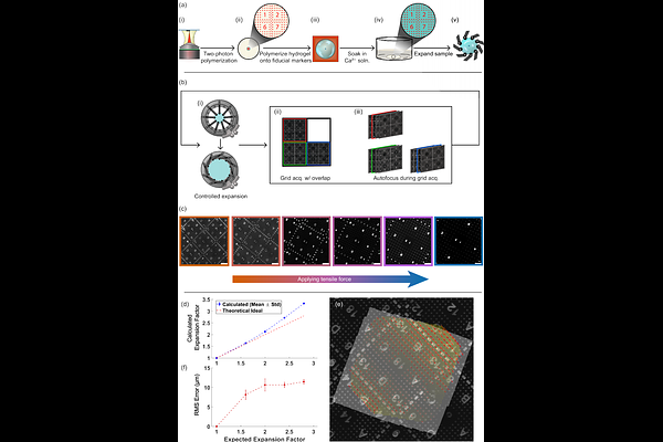
AbstractUnderstanding biophysical phenomena requires techniques that access biologically relevant spatial and temporal scales. Expansion Microscopy (ExM) is a sample preparation approach which achieves super-resolution spatial scales by leveraging osmotic forces in a swellable hydrogel to physically separate structures to distances larger than the diffraction limit of light. Yet, in traditional osmotic ExM only pre- and post-expanded samples can be imaged. Further, fragmentation, hydrogel deformation, and signal loss are common while requiring samples to be chemically fixed. Therefore, there is little control of the expansion, reproducibility can be challenging, and dynamics of biological samples at applicable temporal scales cannot be observed. Here, we develop Tensile Expansion Microscopy (TExM) to mechanically expand fixed and, notably, living cellular samples. Highly-stretchable and tough double network alginate-Ca2+/polyacrylamide hydrogels are expanded by tensile forces applied using an electromechanical iris expansion device during continuous imaging on a fluorescence microscope. We incorporate two-photon polymerized microscale fluorescent fiducial markers to track samples and distortion during expansion. The hydrogels controllably and repeatedly expand up to 3.3x with distortions less than 12 m across 1.3 mm2. TExM is first applied to fixed NIH 3T3 fibroblast cells with immunohistochemistry-stained microtubules, achieving super-resolutions of 100 nm. Then, TExM is demonstrated with living HeLa cells with internal fluorescent reporters showing increased cell size and cell-to-cell separation under 3.2x linear expansion. Overall, TExM allows for continuous, stepwise, and precise temporal modulation of lateral substrate strain, enabling real time monitoring of dynamics of both fixed and viable live cell processes at higher spatial resolutions. TExM can further investigate broad biophysical questions due to its compatibility with other analytical imaging methods that are sensitive to water or fixatives used in traditional osmotic ExM.