Characterizing MINFLUX imaging performance with DNA origami
Characterizing MINFLUX imaging performance with DNA origami
Clowsley, A. H.; Bokhobza, A. F. E.; Janicek, R.; Kołataj, K.; Bleuer, G.; Di Michele, L.; Acuna, G. P.; Soeller, C.
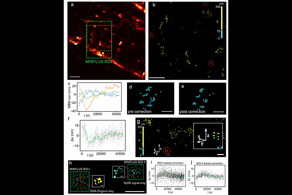
AbstractMINFLUX, a second-generation super-resolution technique, can localize fluorescent markers approaching single-nanometer precision in three dimensions. Similar to previous super-resolution methodologies, the extended duration of acquisitions can result in drift that needs accurate correction to enable proper analysis and interpretation of the collected data. Here, we use DNA origami, housing sites of known spatial distribution fitted with repeat-domain docking strands for DNA-PAINT imaging to characterize imaging performance over extended duration MINFLUX acquisitions (6-20 h). Repeat-domain docking strands overcome site-loss and reveal residual drift in prolonged MINFLUX 3D acquisitions that we correct with an algorithm exploiting time-correlated shifts of localizations around identified DNA origami sites. Following correction of residual drift the site precision, i.e. the scatter of localizations around sites, is ~2 nm in all directions. Comparison of site precision from extended repeat-domain docking strands with site precision from standard short 8-10 nucleotide docking strands exhibits no detectable loss of site precision. By adding DNA origami structures to mounted biological samples we apply our approach to the imaging of the cardiac ryanodine receptor 2 in cryosectioned heart tissue. The data suggests that for these protein targets single domain markers with repeat domain docking strands may be directly used for residual drift correction, simplifying sample preparation and acquisition protocols.