N-cadherin orientational order decreases with mechanical load at cardiomyocyte adherens junctions

N-cadherin orientational order decreases with mechanical load at cardiomyocyte adherens junctions
Tran, Y. T. B.; Dean, W. F.; Han, Y.; Karpov, K. I.; Ainslie, C. M.; Mattheyses, A. L.; Kwiatkowski, A.
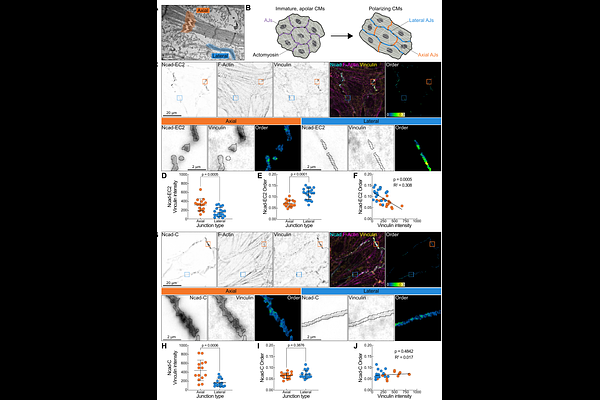
AbstractAdherens junctions physically connect neighboring cells and are built around classical cadherins, homophilic transmembrane proteins that link to the actin cytoskeleton. Classical cadherins can organize into ordered arrays in vitro, but whether they do so in cells remains to be established. Here, we use fluorescence polarization microscopy to show that the classical cadherin N-cadherin is orientationally ordered at cardiomyocyte cell-cell junctions. Whereas the desmosomal cadherin desmoglein 2 was similarly ordered across junction types, N-cadherin order was spatially heterogeneous. Order was lowest where organized myofibrils terminate at high-load, vinculin-enriched axial junctions and highest at low-load, vinculin-poor lateral junctions. This inverse relationship between order and mechanical load suggests that robust cadherin-mediated adhesion does not require ectodomain order. Our findings provide evidence that a classical cadherin is orientationally ordered in cells and show that mechanically active adhesions adopt distinct organizational strategies according to local mechanical demands.