Nanoscale imaging of expanded cells and proteins with spontaneously blinking dyes
Nanoscale imaging of expanded cells and proteins with spontaneously blinking dyes
Sauer, M.; Taban, D.; Jungblut, M.; Budiarta, M.; Helmerich, D. A.; Kiesel, C.; Doose, S.; Kollmannsberger, P.; Plutkiss, S.; Lavis, L.; Rizzoli, S.; Shaib, A. H.; Krah, D.; Beliu, G.
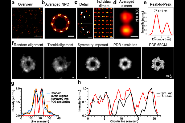
AbstractExpansion microscopy (ExM) enables nanoscale imaging on standard microscopes, but combining ExM with single-molecule localization microscopy (SMLM) remains difficult, owing to the incompatibility of expanded hydrogels with photoswitching buffers. Here, we introduce a single-step expansion microscopy method that allows SMLM with spontaneously blinking dyes in 6-14-fold expanded samples, without re-embedding. We demonstrate nanometer-resolution imaging by resolving the organization of the nuclear pore complex (NPC) and the molecular structure of recombinant homotrimeric proliferating cell nuclear antigen (PCNA).