A green fluorescent protein for live imaging in hyperthermophiles
A green fluorescent protein for live imaging in hyperthermophiles
Kuo, Y.-W.; Radoux-Mergault, A.; Dubois, T.; Cezanne, A.; Zhang, F.; Penttilä, P. A.; Wagner, M.; Dey, G.; Albers, S.-V.; Baum, B.
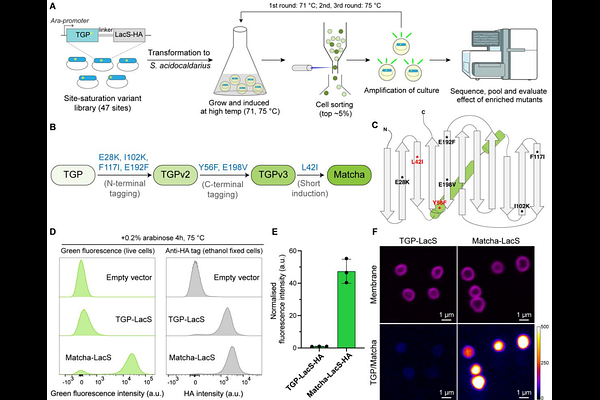
AbstractHyperthermophiles, organisms that thrive at temperatures above 60 {degrees}C, have played important roles in biotechnology and promise to reveal new biology. However, how these cells live remains poorly understood in part due to the lack of bright, thermostable fluorescent proteins that can be used to study protein localisation and dynamics at high temperatures. To overcome this challenge, here we describe the development of ''Matcha'', a green fluorescent protein that we have engineered from Thermal Green Protein by directed evolution in the thermophilic archaeon Sulfolobus acidocaldarius. The screen identified 7 mutations that when combined led to an ~50-fold increase in the brightness of Matcha in vivo at physiological temperatures. Since this is sufficient for live cell imaging, we were then able to use Matcha-fusion proteins to study the division ring dynamics in Sulfolobus. Remarkably, this analysis reveals that, while ESCRT-III rings are disassembled as cells complete division, CdvA forms a stable polymeric ring that persists, and is asymmetrically inherited by one of the two daughter cells following cytokinesis. This study highlights the power of Matcha as a tool to shed light on our understanding of the cell biology of hyperthermophiles.