Quantifying Brittle Crack Opening in Human Trabecular Bone Using Synchrotron XCT-DVC
Quantifying Brittle Crack Opening in Human Trabecular Bone Using Synchrotron XCT-DVC
Vasooja, D.; Cinar, A.; Mostafavi, M.; Marrow, J.; Reinhard, C.; Hansen, U.; Abel, R. L.
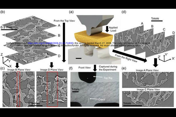
AbstractIntroduction Trabecular bone exhibits brittle behaviour governed by microscale deformation and damage processes, yet quantitative characterisation of crack progression remains challenging because classical fracture-mechanics approaches do not apply to architecturally discontinuous porous tissues. This study evaluates whether synchrotron X-ray computed tomography (XCT) combined with digital volume correlation (DVC) can provide a practical experimental approach for quantifying crack opening behaviour in human trabecular bone. Method Semicylindrical specimens harvested from femoral heads of hip-fracture donors (n = 5) and non-fracture controls (n = 5) underwent stepwise three-point-bending during XCT imaging. Full-field displacement maps enabled direct measurement of crack mouth opening displacement (CMOD), crack length (a), and their ratio, CMOD/a, used here as a geometry-normalised comparative descriptor of brittle response. Automated crack segmentation using phase-congruency crack detection (PCCD) was compared against manual measurements. Results XCT-DVC successfully resolved three-dimensional displacement discontinuities during crack initiation and propagation in all specimens. Hip-fracture donors exhibited significantly lower critical crack-opening ratios (CMOD/a)* than Controls (0.31 vs 0.47; p = 0.008) and reached mechanical instability at lower applied loads, consistent with a more brittle structural response under this test configuration. Despite these differences, total crack extension (da*) was similar between groups. Automated crack tracking using phase-congruency-based segmentation showed excellent agreement with manual measurements (r2 = 0.98), confirming reliable extraction of crack geometry from DVC displacement fields. Discussion These results indicate that XCT-DVC can provide a practical approach for quantifying crack-opening behaviour in trabecular bone when classical fracture-mechanics parameters are not applicable in anatomically constrained specimens. The reduced critical crack-opening ratios and earlier instability observed in Hip-fracture donors are consistent with a more brittle comparative mechanical response that is not captured by crack extension alone. The strong agreement between automated and manual crack measurements further validates displacement-based descriptors as reliable comparative indicators of brittle behaviour in porous, architecturally discontinuous tissues.