Refractive index as an indicator for dynamic protein condensation in cell nuclei
Refractive index as an indicator for dynamic protein condensation in cell nuclei
Marin, O.; Kirchweger, P.; Dalaloyan, A.; Barak, Y.; Elbaum, M.
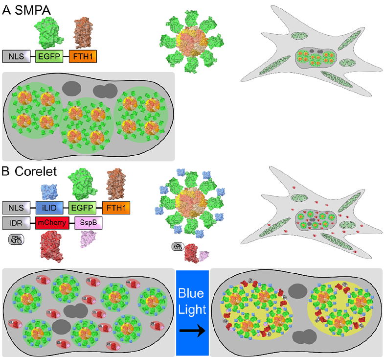
AbstractProtein condensation is the basis for formation of membrane-less organelles in the cell. Most famously, weak, polyvalent interactions, often including RNA, may lead to a liquid-liquid phase separation. This effect greatly enhances local concentrations and is thought to promote interactions that would remain rare in dilute solution. Synthetic systems provide a means to clarify the underlying biophysical mechanisms at play, both in vitro and in the cell via exogenous expression. In this regard, ferritin is a useful substrate, as its composition of 24 subunits with octahedral symmetry supports self-assembly by close packing in 3D. The conventional diagnostic tool for protein condensation is fluorescence imaging. In this work we explore the use of refractive index mapping to detect states of condensation and decondensation. Using two related ferritin-based self-assembly systems, we find that refractive index is a sensitive indicator for reversible condensation. Surprisingly, refractive index indicates a rapid decondensation even when molecular dispersal kinetics are slow according to fluorescence. Conversely, in a photo-activated condensation where long activation results in slow decondensation kinetics, the refractive index provides reliable evidence for the physical state independent of fluorescence. The observations suggest a distinction between condensation to a sparse biomolecular network, or to a material continuum that supports an optical polarizability distinct from that of the dilute phase in solution.